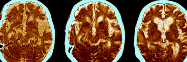
Alzheimer’s disease (AD) remains one of the most perplexing neurodegenerative disorders, affecting millions worldwide. Understanding the molecular underpinnings of this disease is a key step in developing effective interventions. IXRF Systems recently collaborated with Prof. Lee Makowski from Northeastern University to explore the potential of laboratory-based microXRF technology—specifically our Atlas system—to analyze human brain tissues and uncover insights into disease progression. By providing precise elemental mapping capabilities, the Atlas offers new avenues for studying the progression of this complex disease and its molecular hallmarks.
Mapping Neurodegeneration with MicroXRF
MicroXRF has emerged as a powerful analytical tool for visualizing the spatial distribution of chemical elements in biological samples. Unlike traditional staining techniques, microXRF offers nondestructive elemental mapping with high spatial resolution, allowing researchers to identify subtle variations in tissue composition. This capability is crucial for studying Alzheimer’s disease, whose pathological hallmarks include amyloid-β plaques and tau protein tangles. Additionally, its ability to detect elemental distributions without altering the sample’s morphology makes it an invaluable tool in neuropathology.
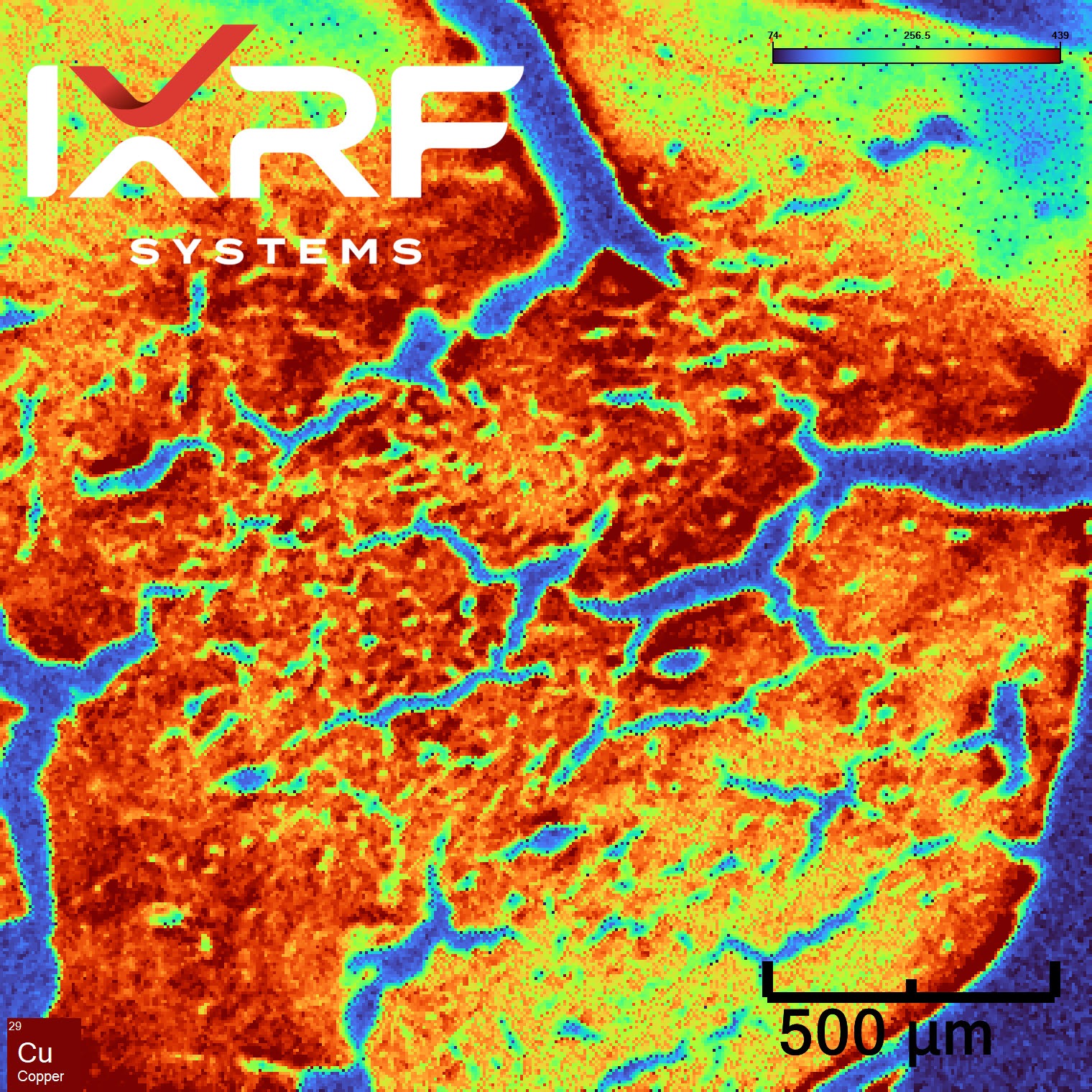
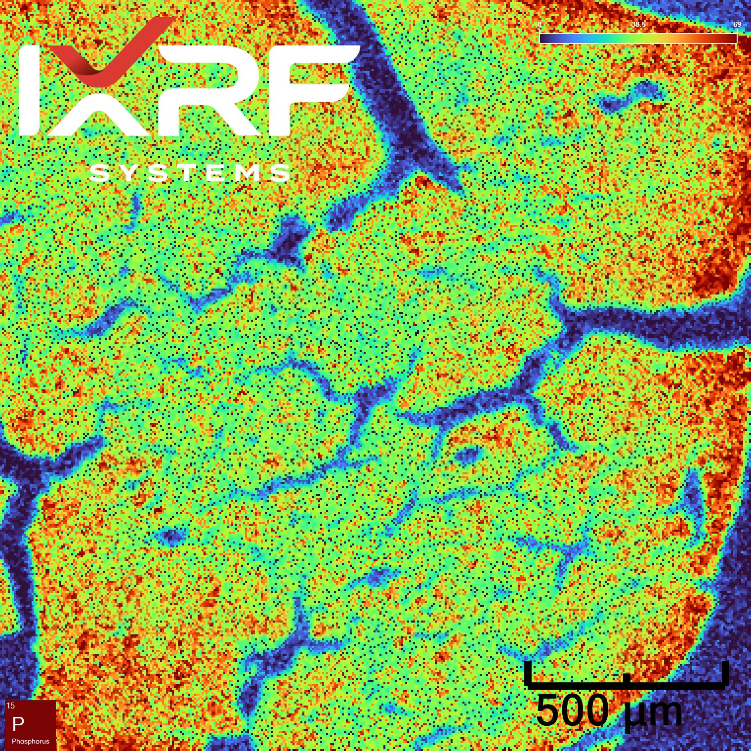
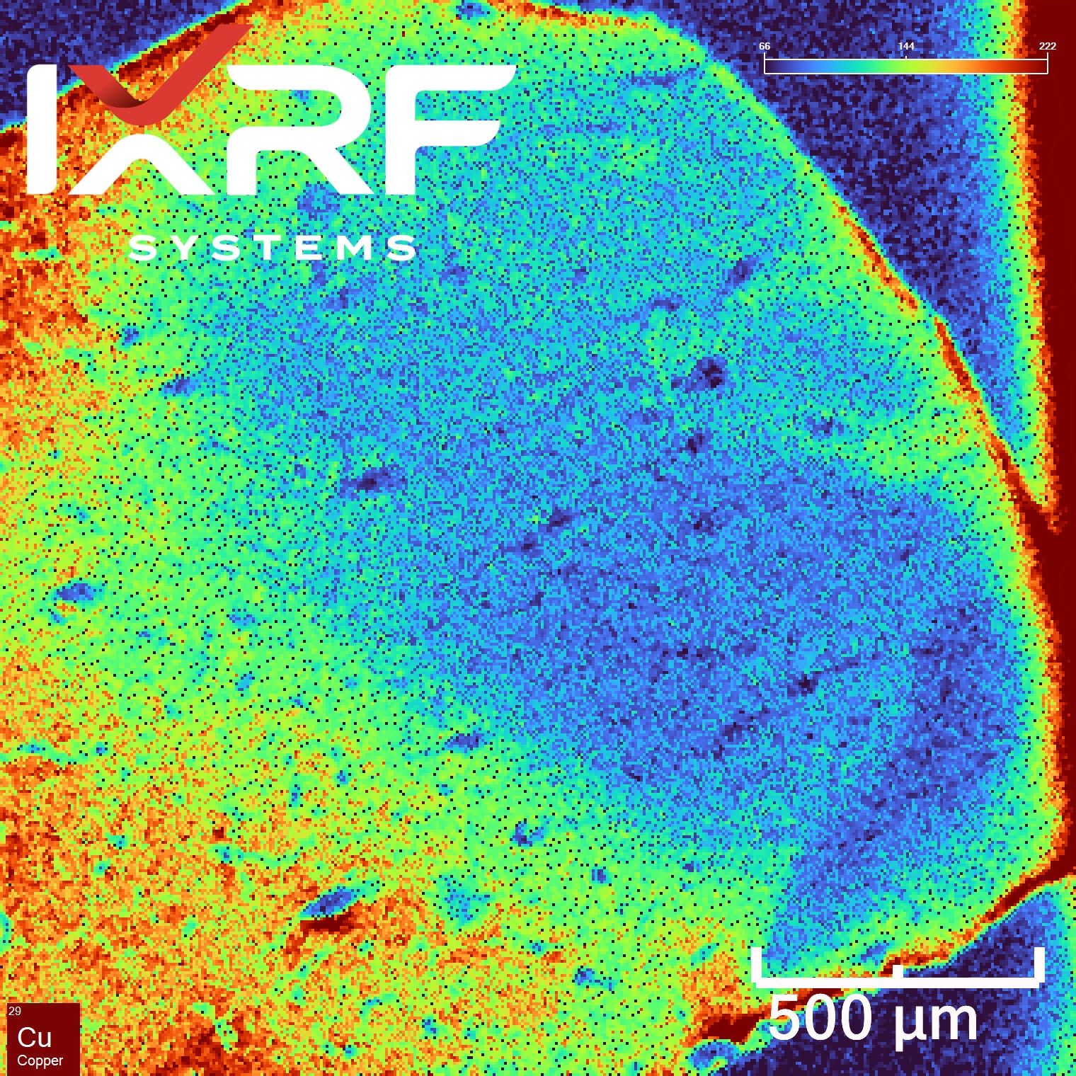
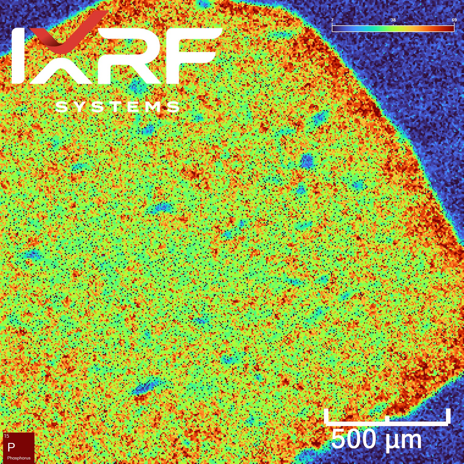
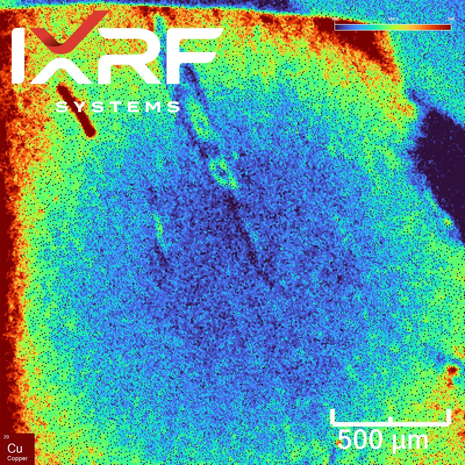
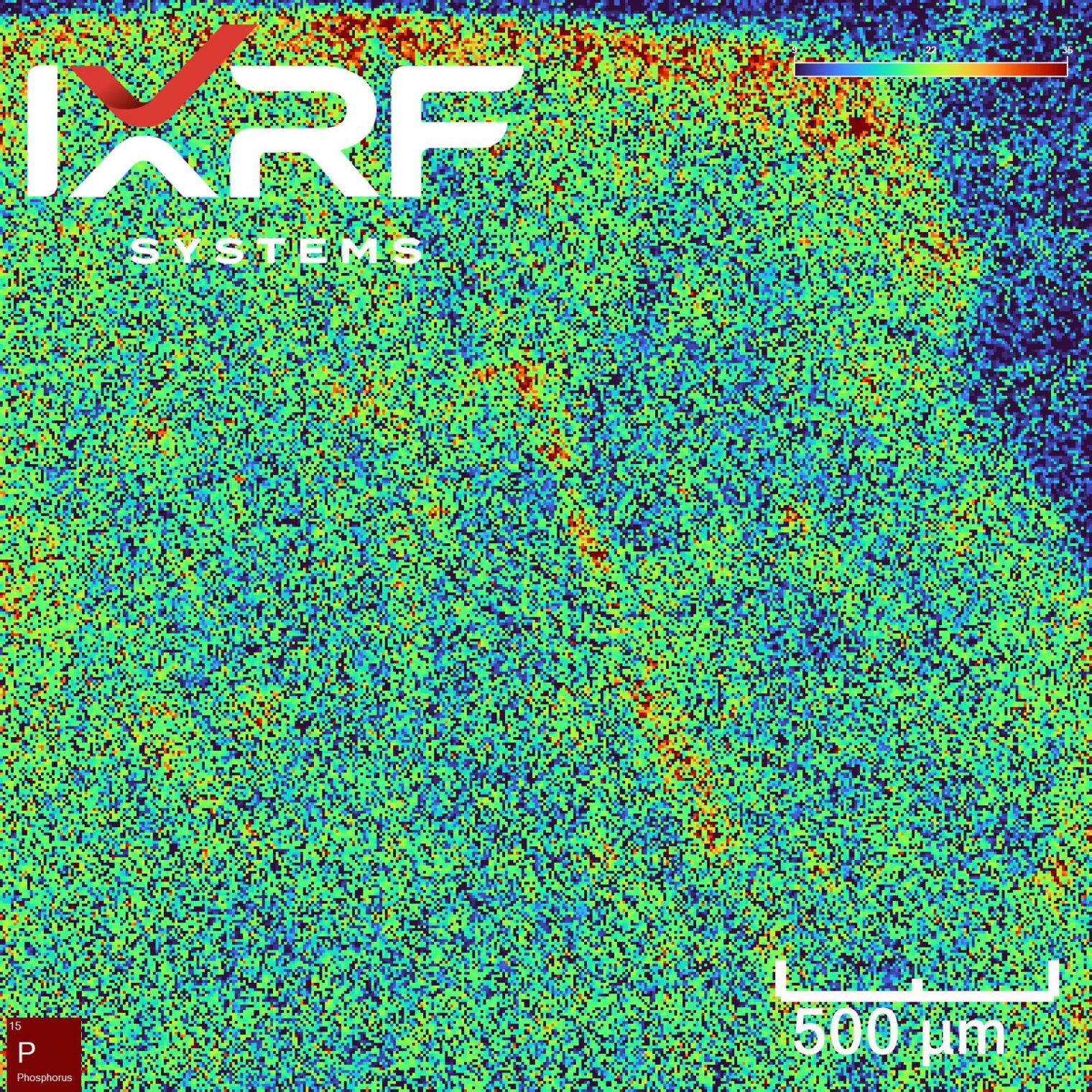
In the study led by Prof. Makowski, our Atlas microXRF was used to map phosphorus (P), iron (Fe), and copper (Cu) within histological thin sections of human brain tissue. Phosphorus, in particular, plays a critical role in cellular processes, and its accumulation near tau protein aggregates may provide clues to disease mechanisms. Previous synchrotron studies, such as those at the European Synchrotron Radiation Facility, highlighted the potential of microXRF for mapping phosphorus at subcellular levels, though access to such facilities is limited. The ability of laboratory-based microXRF to deliver comparable insights makes it a game-changing technology for neuropathological research. Furthermore, the high accessibility of the Atlas system ensures that this cutting-edge analysis is no longer confined to specialized facilities, enabling broader research opportunities.
Key Findings from the Collaboration
The collaborative study between IXRF Systems and Prof. Lee Makowski’s team has provided important insights into the elemental composition of brain tissues affected by Alzheimer’s disease. By utilizing the Atlas microXRF system, we analyzed critical elements, such as phosphorus, with high spatial resolution and accuracy. These findings provide valuable data that could further our understanding of the disease’s progression and its biochemical pathways.
- Phosphorus Enrichment Near Pathological Features: The Atlas revealed increased phosphorus concentrations in regions adjacent to tau fibrils. This aligns with findings from synchrotron-based studies, which also observed phosphorus enrichment in amyloid fibril-dense areas. Phosphorylation of tau is considered a pivotal event in Alzheimer’s progression, and localized phosphorus mapping offers a window into these molecular events. Such elemental gradients provide valuable insight into the biochemical pathways involved in neurodegeneration.
- Iron and Copper as Minor Players: While phosphorus remains the primary focus, preliminary data suggest intriguing distribution patterns for iron and copper. Both elements are implicated in oxidative stress and may play secondary roles in disease pathology. Understanding the interplay of these metals with protein aggregates could shed light on their contributions to neurotoxicity and the oxidative damage observed in AD.
- Correlating Serial Section Data: Serial sections stained for tau and amyloid-β were compared to microXRF maps, revealing correlations between elemental distributions and neuropathological features. This layered approach enhances our understanding of disease mechanisms, as it allows researchers to align molecular data with structural observations, creating a holistic view of disease pathology. Such cross-sectional analyses also aid in validating microXRF data with established histopathological techniques.
Figure 1: MicroXRF images of histological thin section human brain tissues.
Advantages of Laboratory-Based MicroXRF
Traditional synchrotron X-ray fluorescence microscopy (XFM) offers unparalleled resolution but comes with significant logistical challenges, including limited access and the need for specialized facilities. The Atlas microXRF system bridges this gap, providing:
- High Spatial Resolution: 5-micron resolution enables precise mapping of elemental distributions, uncovering features at a scale previously accessible only with synchrotrons.
- Ease of Access: Laboratory-based instruments democratize advanced imaging, allowing routine analyses that complement synchrotron data and enabling continuous research without waiting for beamline access.
- Flexibility in Sample Types: The system accommodates a variety of biological and material science samples, broadening its application scope. From analyzing neural tissues to studying mineral deposits, the versatility of the Atlas expands its utility across disciplines.
- Cost-Effectiveness: The Atlas eliminates the need for costly and time-consuming trips to synchrotron facilities, allowing researchers to conduct high-quality elemental mapping in-house, saving time and resources.
- Operational Efficiency: The Atlas system’s user-friendly design ensures streamlined workflows, enabling researchers to quickly set up, analyze, and interpret results with minimal downtime.
By eliminating barriers associated with synchrotron dependence, the Atlas empowers researchers to conduct high-quality analyses in-house, accelerating the pace of discovery.
Broadening the Horizon of Alzheimer’s Research
The findings from this collaboration emphasize the potential of microXRF to advance Alzheimer’s research. By integrating high-spatial-resolution imaging with neuropathological studies, researchers can explore the molecular drivers of disease in greater detail. For example, identifying phosphorus enrichment near tau fibrils could help pinpoint early stages of disease progression, enabling earlier diagnosis and intervention.
Furthermore, this technique could monitor the effectiveness of experimental therapies aimed at reducing phosphorus accumulation or modifying tau phosphorylation. A hypothetical application might involve longitudinal studies in which treated brain tissues are compared to untreated controls to evaluate elemental changes over time. This would provide a direct, quantitative way to assess therapeutic impact.
As Prof. Makowski’s team demonstrated, laboratory-based microXRF is not merely a substitute for synchrotron studies but a complementary tool that can expand the scope of elemental analysis. The Atlas provides actionable data on phosphorus and other trace elements, opening new doors for studying disease progression and developing targeted therapies.
Moreover, microXRF technology’s accessibility fosters collaboration across research groups. With fewer logistical constraints, scientists from various disciplines can integrate elemental mapping into their workflows, enriching the collective understanding of neurodegenerative diseases.
Looking Forward
IXRF Systems is committed to supporting groundbreaking research in the life sciences. With the Atlas, we aim to empower researchers to explore complex biological phenomena precisely and efficiently. Future studies will focus on refining analytical methodologies, enhancing detection sensitivity for trace elements, and expanding applications of microXRF in neurodegenerative disease research. Our ongoing collaboration with leading researchers like Prof. Makowski highlights the transformative impact of microXRF technology in understanding and combating Alzheimer’s disease.
As we continue to innovate, we look forward to supporting novel investigations that leverage the full potential of microXRF. By integrating elemental imaging with other advanced analytical techniques, we envision a future where researchers can unravel the mysteries of complex diseases with unprecedented clarity.
Join the Conversation. Do you have insights or questions about using microXRF for Alzheimer’s research? Connect with us to learn more about how the Atlas system can support your investigations. Together, we can push the boundaries of scientific discovery and create a brighter future for patients affected by Alzheimer’s and other neurodegenerative diseases.
